Tuesday, March 07, 2006
Genetic lab techniques for antenna-heads
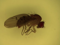
For those wondering how I hunt Wolbachia in insects: I usually start with whole insect specimens, like this fly (genus Drosophila), which I collected from a mushroom bait pile in Rochester, NY last summer.

I store my insects in 95% ethanol, in a freezer at -20ºC. This preserves both the insect tissue and the bacterial cells within. When it's time to isolate DNA from my insects, I use a commercial DNA isolation kit; here's a sample protocol.
When I've ground up enough insects and isolated DNA from them, it's time to screen them for the presence of Wolbachia DNA. To do this, I amplify a Wolbachia gene via the polymerase chain reaction (PCR), using appropriate primers.
Sometimes a DNA isolation fails due to lab error, specimen deterioration, or an unfavorable phase of the moon, so for a DNA quality control, I also amplify an insect mitochondrial DNA (mtDNA) gene, using a different set of primers. The rationale for this is:
* If I can amplify both an insect mtDNA gene and a Wolbachia gene, the insect is infected with Wolbachia.
* If I can amplify an insect mtDNA gene but not a Wolbachia gene, the insect is not infected with Wolbachia.
* If neither gene can be amplified, there's probably something wrong with my DNA isolation or with the reaction I set up.
Since everyone who works in a lab can and will mess up sometimes, I also run negative and positive controls. The positive control is usually a Wolbachia-infected insect. For my Wolbachia gene negative control, I usually use DNA from an insect that I know is uninfected; for my insect mtDNA control, I just add a drop of sterile water to the PCR reaction in place of the insect DNA solution.
I run PCR reactions in 10 microliter volumes, and after I'm done, I load 4 microliters of each reaction onto an agarose electrophoresis gel. I apply about 100 volts to the gel, which drives my amplified DNA fragments through the gel. The gel contains a small amount of ethidium bromide, which makes DNA fluorescent under ultraviolet light. After about a half hour at this voltage, the fragments have migrated far enough to become visible as bands under a UV illuminator.

The lower row of bands on this gel shows the results of my Wolbachia screen on five insects plus positive and negative controls. From left to right:
Lanes 1-3: Infected insects.
Lanes 4-5: Uninfected insects.
Lane 6: Negative control.
Lane 7: Positive control.
Lane 8: Unused.
The upper row shows the results of my screen for insect mtDNA. There are bands present for all five screened insects, showing that the isolated DNA is of good quality. As above, lane 6 is a negative control, lane 7 a positive control, and lane 8 unused.
At this point, I can do something more exciting, like sequencing the Wolbachia and insect DNA. In fact, I'm working on a batch right now, so it's a good place as any to take a break from today's lesson. Just remember: Keep your samples well-separated, your reagents cold, and your fingers away from the ethidium bromide.

I store my insects in 95% ethanol, in a freezer at -20ºC. This preserves both the insect tissue and the bacterial cells within. When it's time to isolate DNA from my insects, I use a commercial DNA isolation kit; here's a sample protocol.
When I've ground up enough insects and isolated DNA from them, it's time to screen them for the presence of Wolbachia DNA. To do this, I amplify a Wolbachia gene via the polymerase chain reaction (PCR), using appropriate primers.
Sometimes a DNA isolation fails due to lab error, specimen deterioration, or an unfavorable phase of the moon, so for a DNA quality control, I also amplify an insect mitochondrial DNA (mtDNA) gene, using a different set of primers. The rationale for this is:
* If I can amplify both an insect mtDNA gene and a Wolbachia gene, the insect is infected with Wolbachia.
* If I can amplify an insect mtDNA gene but not a Wolbachia gene, the insect is not infected with Wolbachia.
* If neither gene can be amplified, there's probably something wrong with my DNA isolation or with the reaction I set up.
Since everyone who works in a lab can and will mess up sometimes, I also run negative and positive controls. The positive control is usually a Wolbachia-infected insect. For my Wolbachia gene negative control, I usually use DNA from an insect that I know is uninfected; for my insect mtDNA control, I just add a drop of sterile water to the PCR reaction in place of the insect DNA solution.
I run PCR reactions in 10 microliter volumes, and after I'm done, I load 4 microliters of each reaction onto an agarose electrophoresis gel. I apply about 100 volts to the gel, which drives my amplified DNA fragments through the gel. The gel contains a small amount of ethidium bromide, which makes DNA fluorescent under ultraviolet light. After about a half hour at this voltage, the fragments have migrated far enough to become visible as bands under a UV illuminator.

The lower row of bands on this gel shows the results of my Wolbachia screen on five insects plus positive and negative controls. From left to right:
Lanes 1-3: Infected insects.
Lanes 4-5: Uninfected insects.
Lane 6: Negative control.
Lane 7: Positive control.
Lane 8: Unused.
The upper row shows the results of my screen for insect mtDNA. There are bands present for all five screened insects, showing that the isolated DNA is of good quality. As above, lane 6 is a negative control, lane 7 a positive control, and lane 8 unused.
At this point, I can do something more exciting, like sequencing the Wolbachia and insect DNA. In fact, I'm working on a batch right now, so it's a good place as any to take a break from today's lesson. Just remember: Keep your samples well-separated, your reagents cold, and your fingers away from the ethidium bromide.